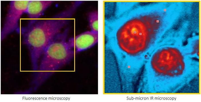

Submicron IR microscopy is opening new opportunities in biological sciences with the development of a new technique: optical photothermal infrared (O-PTIR) spectroscopy.
Image Credit: Photothermal Spectroscopy Corp
Developments in life sciences research are playing an increasingly important role in global health, and advancements in technology have the potential to offer new and improved approaches to support the health and wellbeing of people worldwide.
As these developments emerge, public and private funding for life sciences research increases, enabling researchers to upgrade their analytical and scientific tools with advanced methods, models, and algorithms to drive further scientific discoveries.
Life sciences involve the study of living organisms, including humans, plants, and animals; a deep understanding of the chemical composition of cellular and biomolecular components and processes is essential for advancing life sciences research.
Several methods are available to study these molecular processes and chemical structures, such as fluorescence microscopy, a vital analytical technique that offers high molecular specificity for a range of applications.
However, researchers often face significant limitations when using fluorescence microscopy, especially when they need chemical molecular structural information.
Infrared (IR) and Raman spectroscopy and imaging systems are well-established approaches in life sciences research. Both offer broad macromolecular and spatially resolved characterization features and have been applied across numerous areas of study, including:
- Investigating the biochemical environment within individual cells
- Observing how cells react to drugs
- Processing and quality control in the pharmaceutical industry
However, traditional IR has a restricted spatial resolution for biological research, and Raman can suffer from fluorescence interference, which limits its use in conjunction with fluorescence microscopy.
O-PTIR spectroscopy has overcome many of these limitations and also complements Raman spectroscopy. It can be integrated with fluorescence microscopy, as well.
This article introduces O-PTIR technology, examines several important life sciences applications, and explains how its integration with Raman and fluorescence techniques can benefit life sciences research.
A new approach to IR and Raman spectroscopy and imaging
While there are many benefits to conventional IR and Raman spectroscopy methods, they also have drawbacks that typically prevent their widespread adoption in mainstream life sciences research.
The major limitations of IR spectroscopy include its relatively poor spatial resolution of approximately 10 to 20 micrometers (µm), significant water absorption that can limit live-cell analysis, and susceptibility to spectral distortions from Mie scattering. These distortions can be influenced by the sample’s morphology as well as its chemical composition.
Although Raman spectroscopy is water-compatible and has an excellent spatial resolution of less than one micron, it is susceptible to autofluorescence issues that can mask weak Raman signals.
Raman scattering is also naturally weak, as it often suffers from a low signal-to-noise ratio. This necessitates comparatively long measurement times.
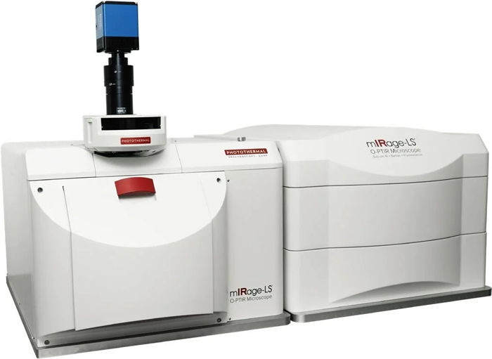
Figure 1. The mIRage-LS Sub-500nm IR multimodal microscope. Image Credit: Photothermal Spectroscopy Corp
However, the introduction of O-PTIR spectroscopy with simultaneous Raman capabilities addresses these challenges directly. O-PTIR can provide sub-micrometer IR spatial resolution without scattering artifacts, is water-compatible, and can simultaneously deliver IR and Raman spectroscopy. This provides the same spatial resolution from the same spot, at the same time.
Researchers can benefit from the chemical complementarities of IR and Raman synergistically for the first time. These methods provide sub-cellular resolution combined with Raman and fluorescence imaging resolution, delivering broad macromolecular characterization on biologically relevant spatial scales below 500 nanometers (nm).
Plus, the integration of widefield epi-fluorescence imaging into the sub-micrometer simultaneous IR and Raman platform, introduced through the mIRage-LS system, allows researchers to combine these techniques into a single instrument without requiring sample registration between measurements, marking a significant leap forward in life sciences research.
The mIRage-LS system allows researchers to take advantage of these two incredibly complementary techniques, which together reveal insights neither technique could uncover on its own.
Plus, the addition of the new counter-propagating mode provides further improvements in IR spatial resolution and extends the range of applications for O-PTIR spectroscopy.
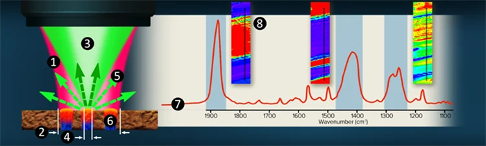
Figure 2. O-PTIR co-propagating technique overview. Image Credit: Photothermal Spectroscopy Corp
Overcoming the IR spatial resolution barrier with O-PTIR
O-PTIR microspectroscopy has removed the 10 to 20 micrometer spatial resolution limitation of IR spectroscopy and enabled IR to be used for sub-micrometer spatial resolution, high-resolution (under 500 nm) chemical structural imaging, and cellular and sub-cellular structure spectroscopy.
The O-PTIR reflection mode co-propagating technique
The technique is illustrated in Figure 2.
- A pulsed, tunable, infrared laser source (pump) is focused through a reflective (Cassegrain) objective
- The infrared beam is focused to 10 to 20 µm (diffraction-limited spot)
- A co-linearly introduced visible laser, typically a 532 nm green Raman excitation laser (probe), is focused through the same objective
- The shorter-wavelength green laser can be focused to a 30-times tighter spot at around 500 nm, allowing it to solely focus on the blue region
- As a function of IR laser wavelength adjustment, the green reflected (or transmitted) laser is detected
- When the infrared wavelength overlaps an absorption band in the sample, energy is absorbed, resulting in a photothermal response and a change in the green reflectivity
- Simultaneously monitoring the green laser reflectivity while tuning the IR source wavelengths creates an infrared spectral response
- Fixed frequency imaging or hyperspectral data sets are accessible for mapping chemical functional group distributions
With submicron IR spatial resolution, infrared spectra are recorded indirectly by detecting a reflected or transmitted 532 nm visible laser. High-quality IR spectra with strong signal-to-noise ratios (SNR) can be collected in just a few seconds.
The O-PTIR counter-propagating technique
Figure 3 demonstrates the addition of a new high-spatial-resolution IR mode called counter-propagating, showing another significant advancement provided by the mIRage-LS platform. The key to achieving increased spatial resolution is using conventional high-power glass objectives as the probe delivery and collection optics.
The co-propagating mode sees the IR pump and visible probe beam focused using all-reflective Cassegrain optics. Using counter-propagating, the two beams are separated, with the IR beam directed via the sample’s underside. This configuration makes it possible to use high-power glass objectives for the delivery and collection of the probe beam.
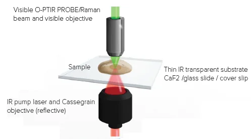
Figure 3. Counter-propagating mode. IR light arrives from the bottom, and the probe laser measures the sample response from the top with a high NA visible objective. Image Credit: Photothermal Spectroscopy Corp
The counter-propagating configuration preserves O-PTIR’s main benefits while providing substantially greater spatial resolution and experimental versatility, including the ability to utilize water-dipping and oil-immersion objectives. Several of these new operating configurations are illustrated in Figure 4.
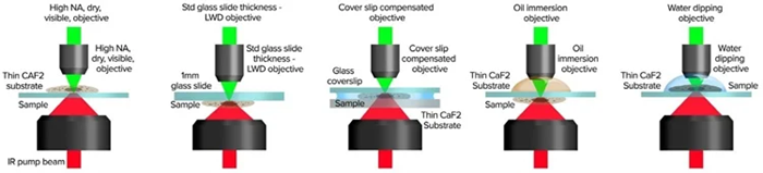
Figure 4. Counter-propagating mode provides application flexibility with a wide range of setups for sample presentation. Image Credit: Photothermal Spectroscopy Corp
Simultaneous Raman and the O-PTIR principle of operation
The unique mIRage-LS design, illustrated in Figure 5, allows Raman and O-PTIR to operate on the same region of the sample simultaneously with identical spatial resolution.
By acquiring both measurements at once, researchers can use the complementary nature of both approaches and provide confirmatory spectral information for more precise results.
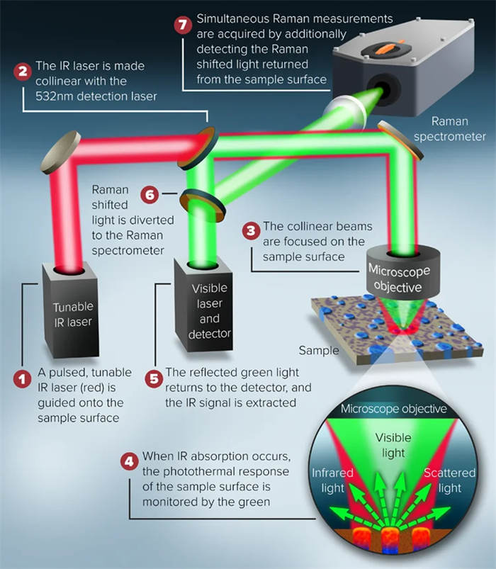
Figure 5. Schematic description of the combined O-PTIR and Raman instrument. Image Credit: Photothermal Spectroscopy Corp
The mIRage LS principle of operation: O-PTIR and fluorescence microscopy
Figure 6 shows the configuration used to combine an O-PTIR submicron IR microscope co-located with fluorescence microscopy. In counter-propagating mode, the pulsed IR beam is focused on the sample’s underside. The fluorescence and visible probe beam paths are co-linear via the microscope objective, allowing measurements to be taken at the same location.
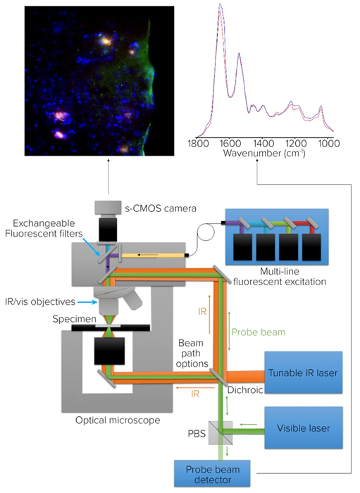
Figure 6. Microscope schematic for O-PTIR and co-located fluorescence microscopy. Image Credit: Photothermal Spectroscopy Corp
O-PTIR uses high-resolution IR spectroscopy and imaging to quantify the same properties that the fluorescence microscope uses to identify biomolecules of interest with high molecular specificity.
To support different fluorescence-based applications, the fluorescence camera incorporates an illumination system not only with a brightfield filter cube, but capacity for seven other filter cubes, too.
Cellular, protein, and tissue applications with O-PTIR and combined techniques
The characterization of neuroglioma cells using counter-propagating mode
Figure 7 shows neuroglioma cells stained with Ras GTPase-activating protein-binding protein 1 (G3BP1) for protein stress granules, 4′,6-diamidino-2-phenylindole (DAPI) for nuclei, and boron–dipyrromethene (BODIPY) for lipids.
In the RGB overlay widefield epifluorescence image on the left, protein stress granules are shown in red, lipids in green, and the nucleus in blue. Square markers indicate the locations where O-PTIR spectra were collected. A brightfield image is on the right.
The O-PTIR spectra obtained from the marked positions in the left- and right-hand panels were collected in just a few seconds and can be seen at the bottom. The measurements reveal distinct spectral differences corresponding to the different sub-cellular regions.
Of particular interest is the slight variation in the amide I band observed in the protein stress granule. This difference indicates a distinct protein secondary structure from the other locations.
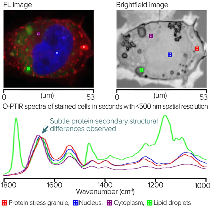
Figure 7. Fluorescence-guided Stained Neuroglioma cells and corresponding O-PTIR spectra. Image Credit: Photothermal Spectroscopy Corp
Monolayer measurement of blood cells using IR and Raman
Figure 8 shows an IR and Raman study of a red blood cell monolayer measured using the copropagating mode from the surface of a glass microscope slide. The image in the left panel covers a 70 by 70 µm area from which a Raman image was collected, with a false color displayed at 1583 cm-1 using 532 nm excitation.
The O-PTIR and Raman spectra shown at the bottom were collected from a selected red blood cell at sub-micrometer spatial resolution and clearly illustrate how effectively IR and Raman spectroscopy work in tandem.
The O-PTIR spectrum is primarily characterized by hemoglobin protein bands, whereas the Raman spectrum is dominated by the heme group’s resonance-enhanced porphyrin ring vibrations.
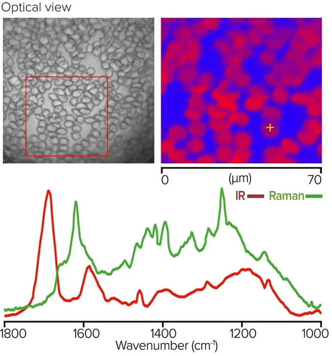
Figure 8. Monolayer of red blood cells, with Raman image and corresponding O-PTIR and Raman spectra. Image Credit: Photothermal Spectroscopy Corp
Tissue analysis with O-PTIR and co-located fluorescence microscopy
Researchers took a brain tissue section from a mouse model of Alzheimer’s disease and analyzed it using Amytracker 630 to identify the amyloid aggregates. AF488 was used to highlight proteins, and DAPI was used to identify the nucleus.
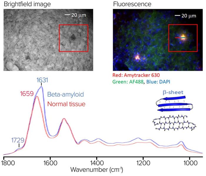
Figure 9. Stained tissue sample with fluorescence image and spectra showing significant spectral differences at 1631 cm-1. Image Credit: Photothermal Spectroscopy Corp
In Figure 9 on the top left, there is a brightfield image of the stained sample. The RGB composite fluorescence image shows amyloid aggregation in red and orange regions. Some of the amyloid aggregates visible through fluorescence are difficult to distinguish in the corresponding brightfield image.
Figure 9 also presents the O-PTIR spectra averaged on and off the aggregate in blue and red, respectively. These spectra were obtained from the line profile marked in the right-hand panel. In the amide I band, where protein beta-sheet structures are characteristic, the aggregate’s average spectrum exhibits various spectral variances and a prominent spectral signature at 1631 cm-1.
This example demonstrates the benefits of combining sub-micrometer O-PTIR spectroscopy with fluorescence imaging. O-PTIR provides molecular compositional information and is particularly sensitive to protein secondary structure, one of the established strengths of IR spectroscopy.
Fluorescence imaging works alongside this to identify regions of amyloid aggregation, some of which are not easily distinguishable using brightfield microscopy alone.
Saltwater algae
Both brightfield and autofluorescence (Ex 650 nm, Em 690 nm) images were captured using a 50x magnification, 0.8 numerical aperture (NA) objective in epi-fluorescence mode.
Figure 10 demonstrates that autofluorescence can display substantial contrast corresponding to known sub-cellular structures compared with the corresponding brightfield image, which contains relatively little structural information.
Because the autofluorescence image provided greater contrast, it was used to direct the subsequent sub-micrometer IR and simultaneous Raman observations.
Although the Raman signals were completely dominated by autofluorescence from the sample, it was still possible to produce high-quality IR spectra without any of the dispersive scattering artifacts typical of direct IR methods such as FT-IR and QCL microscopy. This was done in seconds from spots approximately 500 nm in scale.
To examine the macromolecular distributions further, a simple lipid-to-protein ratio image was generated at 1743 cm-1 and 1655 cm-1 using a single-frequency imaging mode for the lipid and protein.
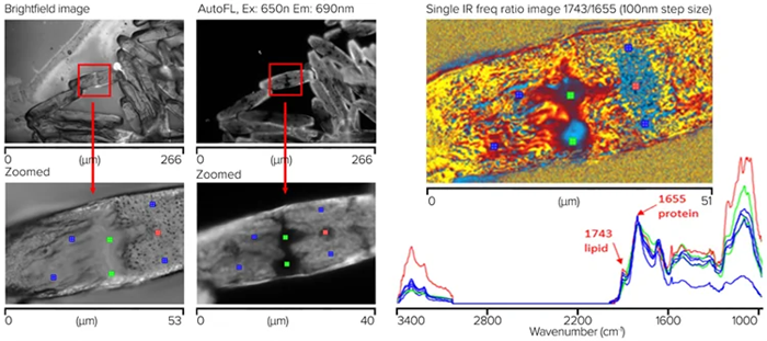
Figure 10. Saltwater algae sample showing measurement locations for the corresponding spectra, with a chemical ratio image (1743/1655 cm-1). Image Credit: Photothermal Spectroscopy Corp
These results demonstrate how this approach could be used by researchers to study such samples against external perturbations, including nutrient changes and depletion and exposure to chemicals and drugs.
Investigating a single bacterial cell using simultaneous O-PTIR and Raman spectroscopy
Simultaneous O-PTIR and Raman spectra were obtained from a single bacterial cell off a CaF2 substrate. A visible image of bacterial cells is shown in the top-left section of Figure 11. The orange box shows where the single-wavenumber IR image at 1655 cm-1 and a 50 nm step size was taken, highlighting the absorption of protein components.
The bottom image shows simultaneous sub-micrometer O-PTIR and Raman spectra taken from a single spot on the single bacterial cell. O-PTIR spectra were acquired in a single unit, using a dual-range QCL covering the 3000–2700 and 1800–950 cm-1 regions. No post-processing was applied to the O-PTIR spectra, but baseline correction was applied to the Raman spectra.
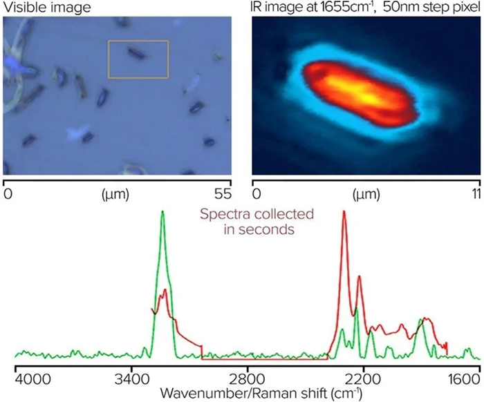
Figure 11. Single bacterial cell, with IR image and corresponding spectra from O-PTIR and Raman. Image Credit: Photothermal Spectroscopy Corp
Fixed cell measurement using an oil immersion objective
To demonstrate the highest levels of optical and IR spatial resolution, researchers used an oil-immersion objective with 100x magnification and 1.3 NA in counter-propagating mode, measuring IR spectra and acquiring images of a human cheek cell on an inverted glass coverslip.
Figure 12 shows how IR features as small as 285 nm can be seen in a rapid ultra-high-resolution image obtained by gathering molecularly specific single-frequency images. In this example, the lipid signal was measured at 1740 cm-1.
Although the cheek cell was mounted on glass, the cellular spectra were not entirely obscured by it, as would be expected with conventional IR methods such as FTIR and QCL microscopy.
Instead, the glass produced a broad silicate feature, over which cellular nucleic acid and carbohydrate signals are observed. Importantly, the protein and lipid regions remain completely free of any contributions originating from the glass.
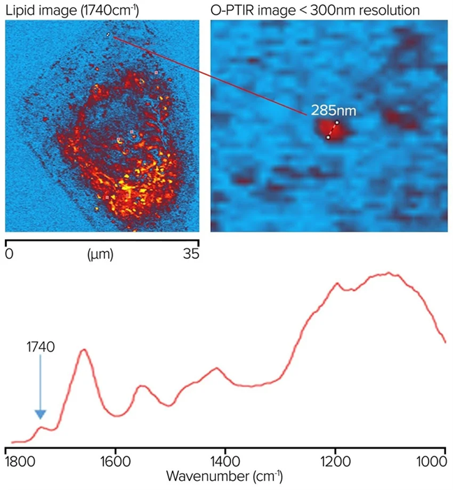
Figure 12. Lipid sample measured at 1740 cm-1 showing a feature resolution of <300 nm with O-PTIR imaging. Image Credit: Photothermal Spectroscopy Corp
Live cells in water using O-PTIR and a water-dipping objective
Cheek cells were produced on a 300 µm-thick CaF2 substrate before researchers applied a drop of water to the sample. A reflecting Cassegrainian objective positioned beneath the sample delivered the IR beam, while a visible water-dipping objective was used to deliver and collect the probe beam in counter-propagating mode, as shown on the right-hand side of Figure 4. A clear image of the sample is displayed in the top-left portion of Figure 13.
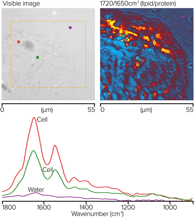
Figure 13. O-PTIR ratio image showing corresponding lipid/protein regions and measured spectra at noted locations. Image Credit: Photothermal Spectroscopy Corp
The orange box shows the region selected for single-wavenumber O-PTIR imaging. Images were collected at 50 nm spacing at 1720 and 1650 cm-1. The top-right corner shows a ratio of these two images. The ratio image’s red and orange parts reflect areas with greater lipid concentrations. Blue sections show higher protein concentrations.
The bottom graphic shows where single-point complete O-PTIR spectra and the appropriately colored spectra were acquired. Although no water background adjustment was used, the spectra show little to no water absorption.
Conclusion
The examples presented in this article demonstrate the range of life sciences applications made possible when using O-PTIR microspectroscopy and imaging in combination with simultaneous Raman and fluorescence measurements.
The mIRage-LS product combines the simultaneous measurement of sub-micrometer O-PTIR and Raman spectra with widefield epi-fluorescence imaging. Bringing these complementary techniques together in a single instrument that offers sample-registration-free operation marks a major breakthrough for life sciences research.
For the first time, researchers can honor established workflows and benefit from the powerful pairing of IR and Raman spectroscopy to access deeper insights that would not be available by using either technique in isolation.
mIRage product overview
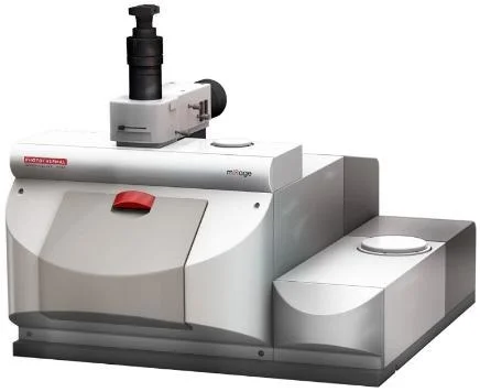
mIRage® – Sub-micron IR spectroscopy. Image Credit: Photothermal Spectroscopy Corp
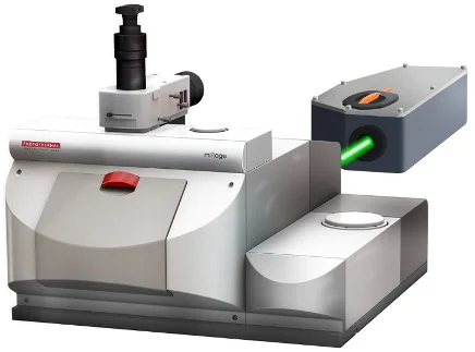
mIRage®-R – Simultaneous sub-micron IR and Raman spectroscopy. Image Credit: Photothermal Spectroscopy Corp
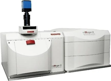
mIRage®-LS – New <500 nm IR with simultaneous Raman and co-located Fluorescence. Image Credit: Photothermal Spectroscopy Corp
About Photothermal Spectroscopy Corp
Photothermal Spectroscopy Corp (PSC) is a Santa Barbara, CA-based scientific equipment company that pioneered sub-micron IR microscopy and spectroscopy. PSC’s vision is to enable the power of sub-micron IR spectroscopy to be applied to high-value problems in both industry and academia via the adoption of O-PTIR and co-located multi-modal techniques.
O-PTIR is one of the highest growth IR microscopy techniques and addresses application areas in life science, microplastics, bioplastics, semiconductors, failure analysis, and chemical and polymeric materials.
mIRage-HSi IR multimodal microscope
mIRage-HSi combines sub-500 nm O-PTIR chemical imaging with laser-scanning speed, revealing label-free distributions of key macromolecules like lipids, proteins and nucleic acids while also resolving micro- and nanoplastic particles in complex biological samples.
Hyperspectral images are acquired in minutes and targeted single-wavenumber images in seconds, making detailed cellular studies and high-throughput particle workflows practical.
The mIRage-HSi supports multi-modal capabilities with simultaneous raman, co-located fluorescence microscopy and optical microscopy making it a powerful instrument for high resolution life science research.
Optical Photothermal IR spectroscopy
Multimodal Optical Photothermal infrared spectroscopy (O-PTIR) used on the mIRage microscope platforms is a result of nearly 2 decades of expertise in photothermal based IR spectroscopy.
O-PTIR: How it works
O-PTIR overcomes the IR diffraction limit by combining a mid-IR pulsed, tunable laser that heats the sample. When the IR laser is at a wavelength that excites a molecular vibration in the sample, absorption occurs, thereby creating photothermal effects including photothermal expansion. A visible probe laser, focused to 0.5 µm spot size, measures the photothermal response via the scattered light, as shown in the illustration above.
The component of the reflected visible laser signal that is modulated at the IR pump laser repetition rate is directly proportional to the absorption coefficient of the sample at that wavenumber. The IR pump laser can be tuned through the entire fingerprint region in one second or less, to obtain an IR spectrum.
By operating in reflection mode, O-PTIR eliminates several longstanding limitations and has substantial benefits for the IR community, including sub-micron resolution using a non-contact optical method. The sub-micron resolution is demonstrated (right), showing reflection mode spectra on a multi-layer packaging film measured 0.5 µm apart with highly differentiated chemical fingerprints indicating different materials.
Measurements are collected quickly and easily without need for sample contact, unlike ATR spectroscopy. Additionally, O-PTIR provides spectra comparable to FTIR without the dispersive artifacts observed in ATR. By operating in reflection mode, the need for thin samples is also eliminated, leading to dramatically easier sample preparation and faster turnaround times.
Sponsored Content Policy: News-Medical.net publishes articles and related content that may be derived from sources where we have existing commercial relationships, provided such content adds value to the core editorial ethos of News-Medical.net, which is to educate and inform site visitors interested in medical research, science, medical devices and treatments.


